Non-invasive temperature tracking with automated infrared thermography: measuring inside out
B.B. Houx 1, M.O.S. Buma 2 and B.M. Spruijt 1
1Animal
Welfare Center, Utrecht University, Utrecht, The Netherlands
2Noldus Information Technology b.v., Wageningen, The Netherlands
Fluctuations in body temperature are frequently used as indicators of an animal's responsiveness to stressors. A well-known short-term response is an immediate increase in core body temperature. On the long term, stressors may affect the amplitude of the circadian temperature rhythm. The two main methods for measuring body temperature are by rectal probes and by telemetry devices. Drawbacks of both methods are (1) that the animal has to be disturbed for the insertion or implantation of the measuring devices, and (2) that the distance between the animal and the receiving apparatus usually limits the study area. Furthermore, changes in body temperature need to be corrected for changes in activity of the animal.
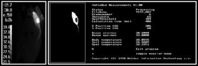
Figure 1. Left: sample of a thermographic recording of a rat. Grey values correspond to temperatures (see index bar on the left). Note the shaven spot (light) on the rat's back. Right: Image after analysis: computer discriminates tail (24.00 °C) and shaven spot (32.34 °C) from the rest of the body (20.06 °C).
We developed a new, non-invasive method for recording body temperature. It combines an infrared thermographic camera with an enhanced version of the automated behavior recording software EthoVision (Noldus Information Technology). The camera outputs a PAL video signal that can be processed by digital imaging software. With this method it is possible to track changes in both behavior and surface temperature continuously (Figure 1). The advantage is that animals can be tracked without disturbing them, in various environments, even in darkness. A new feature is the possibility to measure temperature at different parts of the body (e.g. head and tail) simultaneously (Figure 1, right panel).
The surface temperature measured by our method reflects to a large extent vasoconstriction and -dilation, involved in regulating core temperature. We compared the relationship between both temperatures in rats. On long term (days), skin and telemetrically measured core temperature patterns show a close relationship. On short term (minutes) however, both temperature measures are often at variance. One explanation is that small, autonomously mediated responses to stressors affect skin temperature more directly than core temperature, which may be buffered by other processes. We tested the responsiveness of skin temperature to various stressors in enriched and standard housed rats. Especially the tail temperature response (Figure 2) differed between groups and b etween stressors of different intensity.
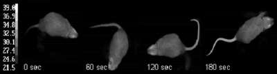
Figure 2. Four samples of a thermographic recording of one rat. Time interval between first and last sample is 180 s. Note the rapid increase of the tail temperature from slightly more than ambient temperature (22 °C) to over 30 °C.
The results indicate that our automated thermographic method may serve as a non-invasive, longitudinal alternative to other methods of temperature measurements. It also provides additional information by detecting small and acute skin (tail) temperature responses to stressors of different severity. We discuss the implications of present method for biophysiological research, its specific limitations and possibilities. We conclude that the method is a useful, additional tool in behavioral, health and welfare research.
Paper presented at Measuring Behavior 2000, 3rd International Conference on Methods and Techniques in Behavioral Research, 15-18 August 2000, Nijmegen, The Netherlands