The SYBAR motion analysis system: integrated recording and display of video, EMG and force-plate data
J. Harlaar, R.A. Redmeijer and P. Tump
Department of Rehabilitation Medicine, University Hospital "Vrije Universiteit", Amsterdam, The Netherlands
In rehabilitation medicine, the assessment of motor dysfunction is an important part of the diagnosis, for indication of therapy. Such assessment includes an analysis of motor disorders by careful observation of the patient who demonstrates his disability or a test of motor performance; e.g. walking. Muscle function can be measured by means of the EMG and total load of the lower leg is represented by the ground reaction force. However, the problem is how to represent these measures in a meaningful way to the physician who is familiar with observational analysis. To do so we developed SYBAR, which is a Dutch acronym for "system for movement analysis in a context of rehabilitation medicine" [1]. SYBAR is based on multimedia technology to integrate digitized video and digitized (physiological) analog signals.
The EMG signal is recorded through a bi-polar leadoff, by two skin electrodes per muscle. This signal is rectified and low-pass filtered (smoothed) at 2.0 Hz. in hardware to obtain the Smoothed Rectified EMG (SR-EMG). The dynamics of SR-EMG approximates the dynamics of muscle force. The SR-EMG signals of several muscles together with six channels of the force-plate (that is immersed in the walkway) are digitized at 100 Hz. These values are stored together with the actual time-code, being the output of time-code generator that is triggered by the video signal. This time-code is also recorded as a part of the video signal, both as a Vertical Interval Time Code (VITC) and inserted as number into the video image. During off-line digitalization of the video, the images are synchronized with the digitized signals. When visualization of the force vector is needed, also a image of a three dimensional calibration objects is made, in order to relate world coordinates to image coordinates.
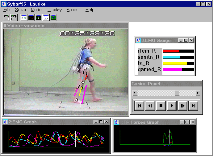
The display of the recordings is obtained by using SYBAR as a viewer (figure 1). User interaction is kept very simple. It employs regular playback and single frame functions. Random access by mouse clicking anywhere in the signal is possible. For a good interpretation of the ground reaction force, it is displayed onto the body (i.e. the video image). In this way, it is possible to estimate joint load for each position.

The SYBAR motion analysis system has been proven very attractive to physicians. The system does not replace the skills of the physician, but extends it by adding explicit information about muscle and joint function. Future developments comprise an integrated analysis of motion (based on feature extraction of the images) and an enhanced integrated online recording of signals and video.

Paper presented at Measuring Behavior '98, 2nd International Conference on Methods and Techniques in Behavioral Research, 18-21 August 1998, Groningen, The Netherlands